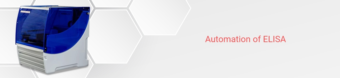
The Gel Doc XR+ System is based on CCD high-resolution, high-sensitivity detection technology and modular options to accommodate a wide range of samples and support multiple detection methods including fluorescence and colorimetric detection. The Gel Doc XR+ System is controlled by Image Lab™ software to optimize imager performance for fast, integrated, and automated image capture and analysis of various samples.
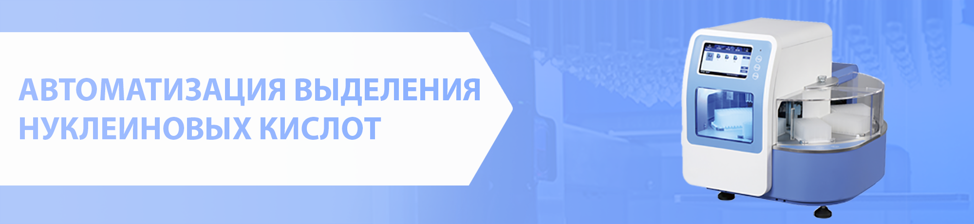
The system accommodates a wide array of samples, from large handcast polyacrylamide gels to small ReadyAgarose™ gels and various blots. The system is an ideal accompaniment to PCR, purification, and electrophoresis systems, enabling image analysis and documentation of restriction digests, amplified nucleic acids, genetic fingerprinting, RFLPs, and protein purification and characterization.
Features and Benefits of the Gel Doc XR+ System
- Gel and blot imaging and analysis are quick and accurate
- Automated, hands-off routines; no training is required
- Save and recall all the steps in the workflow for repeatable and reproducible results
- Optimize the system at setup for image data that is always accurate, reproducible, and free of imaging artifacts
- Wide range of applications with special accessories to preserve sample integrity for downstream research while ensuring user safety
- Comprehensive, automated quantitative analysis of protein and DNA samples in seconds
- Customize and organize data in reports
- Obtain publication-quality results quickly
The Gel Doc XR+ System optimizes reproducibility and reliability of experimental data, enabling quantitative comparisons between different experiments. Using proprietary algorithms, this imaging system is calibrated at setup to ensure:
- Images are in focus at all times, regardless of zoom level or sample position
- Appropriate flat fielding correction is automatically and consistently applied to image data for every application
- Imaging artifacts are automatically corrected
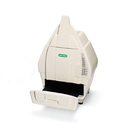
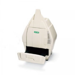
The Gel Doc XR+ System enables quick and easy visualization, documentation, and analysis of nucleic acid and protein gels, blots, and macroarrays with a few clicks of the mouse. The system supports fluorescence and colorimetric detection methods. The Gel Doc XR+ System consists of a darkroom hood, CCD camera and software-controlled motorized lens, UV and white light illuminators, filter slider with standard filter, and UV-protection shield. The system enables you to:
- Increase cloning efficiency and protein production by protecting DNA electrophoresis samples from UV exposure using the XcitaBlue™ conversion screen and blue light excitable stains such as GelGreen, SYBR®Safe, and SYBR® Green I
- Maintain standard operating procedures or criteria for sample performance as there is no loss in sensitivity compared to UV and ethidium bromide staining
- For imaging SYBR Safe DNA applications, you may use the UV transilluminator or the optional XcitaBlue kit. The XcitaBlue (UV to blue conversion screen) protects samples from UV damage and also allows you to externally visualize DNA samples without a UV shield